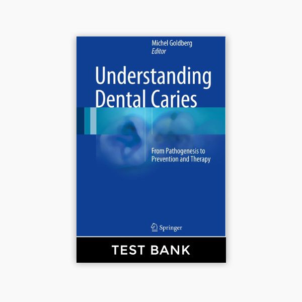
Understanding Dental Caries From Pathogenesis to Prevention and Therapy 1st Edition Goldberg Test Bank is a resource designed to assess knowledge of dental caries, their development, and the prevention and therapeutic interventions to manage them.Focus on the pathogenesis of dental caries, including microbial, dietary, and environmental factors that contribute to their development.Questions related to the clinical and radiographic identification of early and advanced carious lesions. Topics include fluoride application, sealants, dietary counseling, and patient education for caries prevention.
- Part I: The Carious Enamel
- 1: Understanding Dental Caries – from Pathogenesis to Prevention and Therapy
- 1.1 The Carious Enamel
- 1.2 Introduction
- 1.3 Structure
- 1.4 Diagnosis
- 1.5 Which Treatment Is Appropriate (When)
- References
- 2: Enamel Softening (Dental Erosion)
- 2.1 Introductory Remarks
- 2.2 Dental Erosion
- 2.3 Crystallite Dissolution
- 2.3.1 Four Types of Physical and Chemical Dental Erosions Have Been Reported
- 2.3.2 Structure and Chemistry of Dental Erosion
- 2.3.3 Intrinsic Causes of Erosion
- References
- 3: Enamel Etching
- 3.1 The Acid-Etched Enamel: Clinical Usefulness
- 3.2 Effects of Acid or Chelating Solutions on Dental Enamel
- 3.3 The Thickness of Enamel Layers After Etching
- References
- 4: The Early Enamel Carious Lesion
- 4.1 Enamel Structure and Composition
- 4.2 The Early Carious Enamel Lesion
- 4.3 Occlusal and Proximal Caries
- 4.4 Microscopic Changes of Enamel (Figs. 4.4, 4.5, 4.6, and 4.7)
- 4.5 Ultrastructural Changes in Enamel
- References
- 5: Dental Biofilms in Health and Disease
- 5.1 Introduction
- 5.2 Resident Microbiota of the Mouth
- 5.3 Benefits of the Oral Microbiota
- 5.4 Dental Biofilms
- 5.5 Dental Plaque Formation
- 5.6 Microbiology of Caries
- 5.7 Cariogenic Features of Dental Biofilm Bacteria
- 5.8 Theories to Explain the Association of Dental Plaque Biofilms to Caries
- 5.9 Implications for Prevention and Control
- References
- 6: New Caries Diagnostic Methods
- 6.1 Introduction
- 6.2 Laser-Induced Fluorescence Methods
- 6.3 Quantitative Light-Induced Fluorescence (QLF)
- 6.4 Fiber-Optic Transillumination (FOTI)
- 6.5 Near-Infrared Transillumination
- Conclusions
- References
- 7: From the Initial Carious Lesion of Enamel to the Early Development of Coronal Dentin Carious
- 7.1 Dentin Caries: The Different Layers
- 7.1.1 Non-carious Dentin
- 7.1.2 Extracellular Organic Matrix
- 7.2 Size of the Hydroxyapatite Crystallites in the Intertubular Dentin
- 7.3 White and Brown Spots
- References
- Part II: The Carious Dentin
- 8: The Dentinoenamel Junction
- 8.1 Cervical Cemento-Dentinal Formation
- 8.2 Biomechanical Aspects of the Cervical Zone
- 8.2.1 Structural Aspects of Cervical Enamel and Dentin
- 8.2.1.1 Enamel
- 8.2.1.2 Dentin
- 8.2.1.3 Cervical Lesions
- 8.2.2 Mechanical and Tribological Properties of the DEJ
- 8.2.3 Specific Composition of the DEJ
- 8.2.4 Enzymes
- 8.2.5 Abfraction Lesion Formation
- References
- 9: Superficial and Deep Carious Lesions
- 9.1 Different Types of Carious Lesion
- 9.2 Reactionary and Reparative Dentin Formation
- References
- 10: Cervical Sclerotic Dentin: Resin Bonding
- 10.1 Introduction
- 10.2 Noncarious Cervical Sclerotic Dentin
- 10.3 Microstructural Changes in Sclerotic Dentin
- 10.3.1 Tubular Occlusion
- 10.3.2 Hypermineralized Surface Layer in Shiny Sclerotic Lesions
- 10.3.3 Mineral Distribution
- 10.3.4 STEM/EDX Analysis
- 10.3.5 Status of the Collagen Fibrils Within the Surface Hypermineralized Layer
- 10.3.6 Summary of the Microstructural Changes in Noncarious Sclerotic Cervical Dentin
- 10.4 Bonding Adhesive Resins to Sclerotic Dentin
- 10.4.1 Total-Etch Technique
- 10.4.2 Self-Etching Technique
- 10.4.3 Problems in Bonding to Sclerotic Dentin
- 10.4.4 Obstacles in Bonding to Acid-Etched, Sound Dentin
- 10.4.5 Obstacles in Bonding to Acid-Etched Sclerotic Dentin
- 10.4.6 Obstacles to Bonding in Sound Dentin Treated with a Self-Etching Primer Alone
- 10.4.7 Obstacles in Bonding to Self-Etching Primer Treated Sclerotic Dentin
- 10.4.7.1 Sclerotic Dentin with a Thin Hypermineralized Layer (<0.5 μm Thick)
- 10.4.7.2 Sclerotic Dentin with a Thick, Continuous Hypermineralized Layer (>0.5 μm)
- 10.4.8 Summary of Obstacles in Bonding to Sound Versus Sclerotic Dentin
- 10.5 Regional Microtensile Bond Strength Evaluation
- 10.6 Restoring the Class V Sclerotic Lesion
- Conclusions
- References
- 11: The Pulp Reaction Beneath the Carious Lesion
- 11.1 Extracellular Matrix of Sound Pulp: Composition
- 11.2 Cells
- 11.3 Neuropeptides in Dental Pulp
- 11.4 Human T-Lymphocyte Subpopulation
- 11.5 The Carious Pulp
- 11.6 Reactionary and Reparative Dentin Formation
- 11.6.1 Reactionary Dentin Formation
- 11.6.2 Reparative Dentin Formation
- 11.7 Pulp Capping
- References
- Part III: Cervical Erosions
- 12: Ultrastructure of the Enamel-Cementum Junction
- 12.1 Procollagen Fibrillogenesis in Dentinogenesis
- 12.2 Enamel Surface
- 12.3 Morphology of the Cemento-Enamel Junction
- 12.4 Erosive Tooth Wear (Figs. 12.4 and 12.5)
- 12.5 Root Caries
- References
- 13: Cervical Erosions: Morphology and Restoration of Cervical Erosions
- 13.1 Morphology of Cervical Dentin Erosion
- 13.2 Restoration of Cervical Dentin Erosions
- References
- 14: Cervical Regeneration
- 14.1 Abfraction: Non-carious Cervical Lesions
- 14.2 Pulpal Regeneration
- 14.3 Dental Pulp Engineering and Regeneration: Advancement and Challenge
- 14.4 Tooth Resorption
- 14.5 Cervical Regeneration
- References
- Part IV: Fluoride
- 15: Fluoride
- 15.1 Dental Fluorosis
- 15.1.1 Mechanisms of Enamel Fluorosis
- 15.1.2 Fluoride Effects on Dentin Formation
- 15.2 Fluoride in Food and Water
- 15.3 Fluoride and Remineralizing Agents
- 15.3.1 Fluoride Delivered from Oral Care Products
- 15.3.2 Other Remineralizing Agents
- References
- Part V: Invasive and Non-invasive Therapies
- 16: Brushing, Toothpastes, Salivation, and Remineralization
- 16.1 Toothbrushing and Toothpaste
- 16.1.1 Changes in Fluoride Concentration of Saliva During Brushing
- 16.1.2 Modern Toothpastes
- 16.1.3 Modern Toothpaste Components
- 16.1.4 Fluoride and Non-fluoride Mineralization Systems
- 16.1.5 Toothpastes and Abrasivity
- 16.1.6 Toothpastes and Abrasivity Testing
- 16.2 Toothbrush: Manual vs. Power
- 16.2.1 Brushing Techniques
- 16.2.2 Clinical Brushing Studies
- 16.2.3 Power Brush Options
- 16.3 Saliva and Saliva Substitutes
- References
- 17: Resin Infiltration Treatment for Caries Lesions
- 17.1 Background
- 17.2 Uptake of Extraneous Materials into Carious Lesions
- 17.3 Infiltration of Carious Enamel with Resin Materials
- 17.3.1 Background
- 17.3.2 Fissure Sealing
- 17.3.3 Infiltration
- 17.3.4 Resins for Infiltration
- 17.3.4.1 Resorcinol Formaldehyde
- 17.3.4.2 Methacrylate-Based Resins
- 17.3.5 Detection of Resin Within the Lesion and Measurement of Occluded Volume
- 17.3.5.1 Detection of Infiltrated Resin
- 17.3.5.2 Determination of Occluded Pore Volume
- 17.3.6 Effects of Infiltration on Carious Tissue
- 17.3.7 Application to Other Enamel Dysplasias Exhibiting Abnormal Porosity
- 17.3.8 Future Developments
- 17.3.8.1 Design of Infiltration Materials
- References
- 18: Minimally Invasive Therapy: Keeping Treated Teeth Functional for Life
- 18.1 Introduction
- 18.2 Minimal Intervention Dentistry
- 18.2.1 Its Rationale
- 18.2.2 Its Cornerstones
- 18.3 Managing Dental Caries
- 18.3.1 Managing Enamel Carious Lesions: Therapies Other Than Fluoride and Infiltration
- 18.3.1.1 Casein Phosphopeptides-�Amorphous Calcium Phosphate
- 18.3.1.2 Ozone
- 18.3.1.3 Chlorhexidine-Containing Agents
- 18.3.1.4 Sealants
- Indications for Placing a Sealant
- Resin-Based Sealants
- Atraumatic Restorative Treatment (ART) High-Viscosity Glass-Ionomers
- Effectiveness of Fissure Sealants
- Comparison Between ART Sealants and Resin Sealants
- 18.4 Managing Dentine Carious Lesions
- 18.4.1 How Does One Manage a Dentine Carious Lesion?
- 18.4.2 Non-cavitated Dentine Carious Lesions
- 18.4.3 Small Cavitated Dentine Carious Lesions
- 18.4.4 Obvious Cavitated Dentine Carious Lesions in Primary Teeth
- 18.4.4.1 Cleansable Cavitated Dentine Carious Lesions
- Ultraconservative Treatment
- Silver Diamine Fluoride
- Hall Technique
- 18.4.5 Restorative Management of Carious Lesions
- 18.4.5.1 Principles for the Removal of Demineralised Carious Dentine
- 18.4.5.2 How Much Demineralised Carious Dentine Needs to be Removed?
- 18.4.5.3 Which Dentine Carious Tissue Removal Method Is Preferable?
- 18.4.5.4 Disinfecting Excavated Tooth Cavities
- 18.4.5.5 Cavity Lining
- 18.4.5.6 Restorative Materials
- 18.4.5.7 Restoring a Cleaned Cavity
- 18.4.5.8 The Atraumatic Restorative Treatment
- 18.5 How to Manage a Defective Restoration
- 18.6 Concluding Remarks
- References
- 19: Invasive and Noninvasive Therapies
- 19.1 Prevention in Dental Practice
- 19.1.1 What Is Prevention?
- 19.1.2 Individual Caries Risk Assessment
- 19.1.3 Strategies for Prevention
- 19.1.3.1 Education
- Preventing Early Colonization
- Toothbrushing
- Dietary Counseling
- 19.1.3.2 Dental Sealants
- 19.1.3.3 Fluoridated Agents
- Fluoride Toothpaste
- Fluoride Varnish
- Fluoride Mouthwashes
- Fluoride Gels
- Comparison of the Caries-Preventive Effectiveness of One Form of Topical Fluoride Intervention w
- Slow-Release Fluoride Devices
- Fluoride Tablets, Drops, Chewing Gums, and Lozenges
- 19.1.3.4 Non-fluoridated Agents
- Xylitol-Containing Products
- Chlorhexidine
- CPP-ACP
- Other Molecules
- 19.1.4 Follow-Up
- 19.1.5 Caries Management by Risk Assessment
- 19.1.6 Key Messages for Dental Practitioners


Reviews
There are no reviews yet.